Ear
|
|
 https://classroomclipart.com Classroom Clip Art" width="180" height="135" longdesc="/encyclopedia/index.php/Image:Ear.jpg" />
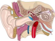
 An ear is an organ used by an animal to detect sound. The term may refer to the entire system responsible for collection and early processing of sound (the beginning of the auditory system), or merely the externally-visible part. Not all animals have ears in the same part of the body.
| Contents |
The ear
Mammals, including humans, have two ears, one on each side of the head.
The Outer Ear
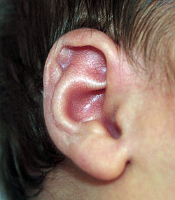
The outer ear is the external portion of the ear. The visible part is called the pinna, or auricle, and functions to collect and focus sound waves. Many mammals can move the pinna in order to focus their hearing in a certain direction, in much the same way that they can turn their eyes. Humans have generally lost this ability. From the pinna, the sound pressure waves move into the ear canal, a simple tube running to the middle ear. This tube amplifies frequencies in the range 3 kHz to 12 kHz.
The human ear has ear lobes at the bottom which are vestigial, but serve the purpose of providing an attachment point for earrings. The earlobe is usually formed cleft from the side of the face and hangs from the rest of the ear, but occasionally will be found looking fused and "lobeless". The helix is the outer edge of the outer ear[1] (http://www.bartleby.com/61/31/H0133100.html).
The Middle Ear
The middle ear includes the eardrum (tympanum or tympanic membrane) and the ossicles, three tiny bones of the middle ear. Their Latin names are the malleus, incus, and stapes, but they are also referred to by their English translations: the hammer, anvil, and stirrup respectively.
Mammals are unique in having three ear bones. The incus and stapes are derived from bones of the jaw, and allow finer detection of sound.
These bones form the linkage between the tympanic membrane and the oval window that leads to the inner ear. The tympanum turns vibrations of air in the ear canal into vibrations of the ossicles. The ossicles in turn transmit the vibrations through the membrane of the oval window into the fluid of the inner ear. The ratio in area between the tympanic membrane and the oval window results in an effective amplication of approximately 14 dB, peaking at a frequency of around 1 kHz. The combined transfer function of the outer ear and middle ear gives humans a peak sensitivity to frequencies between 1 kHz and 3 kHz. The tensor tympani muscle and stapedius muscle of the inner ear contract in response to loud sounds, reducing the transmission of sound to the inner ear. This is called the acoustic reflex.
The middle ear is hollow. If the animal moves to a high-altitude environment, or dives into the water, there will be a pressure difference between the middle ear and the outside environment. This pressure will pose a risk of bursting or otherwise damaging the tympanum if it is not relieved. This is one of the functions of the Eustachian tubes - evolutionary descendants of the gills - which connect the middle ear to the nasopharynx. The Eustachian tubes are normally pinched off at the nose end, to prevent being clogged with phlegm, but they may be opened by lowering and protruding the jaw; this is why yawning helps relieve the pressure felt in the ears when on board an aircraft.
The Inner Ear
The inner ear comprises both the organ of hearing (the cochlea) and the labyrinth or vestibular apparatus, the organ of balance located in the inner ear that consists of three semicircular canals and the vestibule.
The cochlea is a hollow organ filled with endolymph, a fluid medium that receives the sound vibrations transmitted from the air to the oval window through the ear drum and ossicles of the middle ear (see above). The cochlea is wrapped in a spiral shape and also contains the coiled basilar membrane, which resonates preferentially in different locations along its length depending on the frequency of the impinging vibrations. Sitting on top of the basilar membrane is a cellular layer known as the Organ of Corti, which is lined with hair cells - sensory cells topped with hair-like structures called stereocilia. When a region of the basilar membrane resonates, the hair cells in that region send nerve impulses to the brain, which are perceived as a sound of whatever pitch the hair cell is associated with. A very strong movement of the endolymph due to very loud noise may cause hair cells to die. This is a common cause of partial hearing loss, and the reason why anyone near guns or heavy machinery should wear earmuffs or earplugs.
The vestibular apparatus is filled with the same endolymph as the cochlea, but instead of detecting sound, it detects rotation of the head. If a line is drawn through the middle of each of the three semicircular canals, perpendicular to the plane in which the canal lies, the three lines would be perpendicular. They would represent three axes of rotation. Any rotation could be represented as three simultaneous rotations about the three axes.
Anatomy Clipart and Pictures
- Clip Art (https://classroomclipart.com)
- Anatomy Clipart (https://classroomclipart.com/image/category/anatomy-clipart.htm)
- Anatomy Pictures and Illustrations (https://classroomclipart.com/image/category/anatomy-illustrations.htm)
Diseases and medical conditions of the ear and auditory system
Problems with the ear or auditory processing system in the brain can lead to deafness.
- Acoustic neurinoma
- Balance disorders
- Barotrauma
- Benign Paroxysmal Positional Vertigo
- Cholesteatoma
- Ear infections
- Conductive hearing impairment
- Labyrinthine hydrops
- Labyrinthitis
- Meningitis
- Neurofibromatosis Type 1
- Neurofibromatosis Type 2
- Noise-induced hearing loss
- Nonsyndromic hereditary hearing impairment
- Otitis externa
- Otitis media
- Otosclerosis
- Perilymph fistula
- Presbycusis
- Sensorineural hearing loss
- Sudden deafness
- Tinnitus
- Usher syndrome
- Vestibular neuronitis
| Sensory system - Auditory system | Edit (http://en.wikipedia.org/w/wiki.phtml?title=Template:Auditory_system&action=edit) |
| Pinna - Ear canal - Eardrum - Ossicles - Cochlea - Basilar membrane - Organ of Corti - Hair cells - Auditory nerve - Primary auditory cortex |

