Western blot
|
|
Western_blot.jpg
A Western blot is a method in molecular biology to detect a certain protein in a sample by using antibody specific to that protein. It also gives information about the size of that protein. Its name is a pun off the name Southern blot, a technique for DNA detection developed earlier by Edward Southern. Detection of RNA is termed Northern blotting.
Steps in a Western blot
- Gel electrophoresis
The first step is gel electrophoresis. The proteins of the sample are separated according to size on a gel, usually using SDS-PAGE. Usually the gel has several lanes so that several samples can be tested simultaneously. However, it is also possible to use a 2-D gel which spreads the proteins from a single sample out in two dimensions.
- Nitrocellulose transfer
The proteins in the gel are then transferred onto a membrane made of nitrocellulose or PVDF, by pressure or by applying a current. This is the actual blotting process and is necessary in order to expose the proteins to antibody (see below). The membrane is "sticky" and binds proteins non-specifically (i.e. binds all proteins equally well). Protein binding is based upon hydrophobic interactions as well as charged interactions between the membrane and protein.
PVDF is often used because it is sturdier and can be "stripped" of antibodies and reused. Unlike nitrocellulose, PVDF must be soaked in 100% methanol before using.
- Blocking
The membrane is then blocked, in order to prevent non-specific protein interactions between the membrane and the antibody protein (next step, below). This is done by placing the membrane in a solution of Bovine serum albumin (BSA) or non-fat dry milk. Without the blocking, the antibody to be applied in the next step would bind to the nitrocellulose.
- Primary Antibody
The first antibody (often called the primary antibody) is incubated with the membrane. The antibody is diluted in a solution containing a modest amount of a salt such as sodium chloride, some protein (such as BSA) to prevent non-specific binding of the antibody to surfaces and a small amount of a buffer to keep the solution near neutral pH. The diluted antibody solution and the membrane can be sealed in a plastic bag and gently agitated for an "incubation" of about half an hour. The primary antibody recognizes only the protein of interest, and will not bind any of the other proteins on the membrane. It is obtained by immunizing an animal (usually a rabbit or goat) with the protein of interest (i.e., injecting the protein into the animal's body) and collecting the antibodies the animal produces against that protein. Some high affinity monoclonal antibodies can also be used for Western blots.
- Secondary Antibody
After rinsing the membrane to remove unbound primary antibody a secondary antibody is incubated with the membrane. It binds to the first antibody, and is usually produced by a different animal. For example, goat anti-rabbit antibody might be used if the first antibody was produced by rabbits. This secondary antibody is usually linked to an enzyme that can allow for visual identification of where on the membrane it has bound. As with the ELISPOT and ELISA procedures, the enzyme can be provided with a substrate molecule that will be converted by the enzyme to a colored reaction product that will be visible on the membrane (see the figure below with blue bands). Alternately, the reaction product may produce enough fluorescence to expose a sensitive sheet of film when it is placed against the membrane.
An alternative to using an enzyme that is coupled to the secondary antibody is to use a radioactive label. An antibody-binding protein such as Staphylococcus Protein A can be used and labeled with a radioactive isotope of iodine.
- Developing
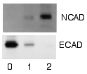
The unbound secondary antibodies are washed away, and the enzyme substrate is incubated with the membrane so that the positions of membrane-bound secondary antibodies will become visible. If a radioactive label is used, the radioactive membrane can be placed against a sheet of medical X-ray film. Bands corresponding to the detected protein of interest will appear as dark regions on the developed film (see figure to right).
Since the first antibody only recognizes the protein of interest, and the second antibody only recognizes the first antibody, if there is stain present on the membrane then the protein of interest must also be present on the membrane. Thus, the protein bands on the membrane that are stained contain the protein that was to be detected, the other locations on the membrane do not. Size approximations can be done by comparing the stained bands to that of a pre-stained protein size marker.
Usually, the gel is not completely devoid of proteins after blotting. Protein staining solution will show all protein bands on the gel. The stained gel can then be compared with the stained membrane to identify which bands contain the wanted protein and which do not.
In principle, one could bind the chemical signal directly to the first antibody, but production of the antibodies is easier if the two functions recognition and signalling are separated.
Medical diagnostic applications
- The HIV test known as "Western Blot" uses a variant of the technique, where the goal is to detect the presence of antibody in a sample. Known HIV infected cells are opened and their proteins separated and blotted on a membrane as above. Then the serum to be tested is applied. Free antibody is washed away, and a secondary antibody is added that binds to human antibody and is linked to an enzyme signal. The stained bands then indictate the proteins to which the patient's serum contains antibody.
- Western blot is also used as the definitive test for BSE or mad cow disease.
External Link
- Western Blot protocols and information (http://www.westernblotting.org)de:Western Blot

