Thalamus
|
|
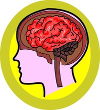
The thalamus is a part of the brain. It is located in the center of the brain, beneath the cerebral hemispheres and next to the third ventricle. It is formed of grey matter and can be thought of as a relay station for nerve impulses in the brain.
| Contents |
Anatomy
The thalami are prominent bulb-shaped regions, typically slightly more than than 1 cm in length, located in the center of the brain. They form on each side of the paleopalliar diencephalon that emerges from the mammalian prosencephalon. The thalamus is largely made of nuclear groups that relate to specific functions in the cerebrum. Thalamic nuclei have subcortical projections, and can be classified as either relay nuclei or association nuclei (see below). Thalamic nuclei also have strong reciprocal connections with the cerebral cortex, forming cortico-thalamo-cortical recurrent loops that are believed may be involved with consciousness.
Function
Most textbooks will tell you that the thalamus is a "relay" that simply relays signals from auditory, somatic, visceral and visual regions of the peripheral nervous system to the cerebral cortex. The real picture is more complicated, and the commonly accepted function of the thalamus nowadays is that it modulates, in addition to relaying, sensory signals to and from cortex.
It is common to classify thalamic nuclei as either "relay nuclei" or "association nuclei" on the basis of the source of their driving inputs, whether they are subcortical or cortical. Relay nuclei receive their driving inputs from subcortical sources including ascending afferents (medial lemniscus for somatosensory information, optic tract for visual information, etc...) and project predominantly to primary sensory cortical areas. On the other hand, association nuclei receive their driving inputs from other cortical areas. (See Sherman and Guillery's "Exploring the Thalamus", 2002)
Alternative ways for subdividing thalamus are also coming into vogue. For example, Ted Jones has recently proposed a Matrix-Core model for the thalamus, which is not subdivided based on nuclei, but rather on chemically-defined populations of neurons. Specifically, Jones proposes the calbindin immunopositive neurons constitute a "matrix", whereas parvalbumin immunopositive neurons form the "core". In Jones' scheme, the "matrix", which includes much of the intralaminar nuclei, is involved in state functions (arousal, level of attention, mood), whereas the "core" is involved in discriminative sensori-motor functions.
Pathology
Cerebrovascular accidents or strokes can cause thalamic syndrome, which results in a burning or aching sensation on one half of a body, often accompanied by mood swings.
Anatomy Clipart and Pictures
- Clip Art (https://classroomclipart.com)
- Anatomy Illustrations (https://classroomclipart.com/clipart/Illustrations/Anatomy.htm)
- Anatomy Clipart (https://classroomclipart.com/clipart/Anatomy.htm)
- Anatomy Animations (http://classroomclipart.com/cgi-bin/kids/imageFolio.cgi?direct=Animations/Anatomy)


