Lung
|
|
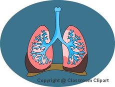
The lung is an organ belonging to the respiratory system and interfacing to the circulatory system of air-breathing vertebrates. Its function is to exchange oxygen from air with carbon dioxide from blood. The process in which this happens is called "external respiration" or breathing. The mechanism by which lungs enable respiration is called diffusion. There are also nonrespiratory functions of the lungs. Medical terms related to the lung often start in pulmo- from the Latin word pulmones for lungs.
| Contents |
Nonrespiratory functions of the lungs
In addition to respiratory functions such as gas exchange and regulation of hydrogen ion concentration, the lungs also:
- influence on the concentration of biologically active substances and drugs used in medicine in arterial blood
- filter out small blood clots formed in the systemic veins
- serve as a physical layer of soft, shock-absorbent protection for the heart, which the lungs flank and nearly enclose.
Mammalian lungs
The lungs of mammals have a spongy texture and are honeycombed with epithelium having a much larger surface area in total than the outer surface area of the lung itself. The lungs of humans are typical of this type of lung. The environment of the lung is very moist, which makes them a hospitable environment for bacteria. Many respiratory illnesses are the result of bacterial or viral infection of the lungs.
Breathing is largely driven by the diaphragm below, a muscle that by contracting expands the cavity in which the lung is enclosed. The rib cage itself is also able to expand and contract to some degree.
As a result, air is sucked into and pushed out of the lungs through the trachea and the bronchial tubes or bronchi; these branch out and end in alveoli which are tiny sacs surrounded by capillaries filled with blood. Here oxygen from the air diffuses into the blood, where it is carried by hemoglobin.
The deoxygenated blood from the heart reaches the lungs via the pulmonary artery and, after having been oxygenated, returns via the pulmonary veins.
Location
The lungs are located inside the thoracic cavity, protected by the bony structure of the rib cage and enclosed by a double-walled sac called pleura. The inner layer of the sac adheres tightly to the outside of the lungs and the outer layer is attached to the wall of the chest cavity. The two layers are separated by a thin space called the pleural cavity that is filled with pleural fluid; this allows the inner and outer layers to slide over each other, and prevents them from being separated easily. The left lung is smaller than the right one to give way for the heart.
Avian lungs
Birds have a complex but highly efficient crosscurrent exchange system in their lungs, accompanied by air sacs to control the flow of gas through it. See the article on bird respiration for a detailed account of this system.
The lungs of birds differ significantly from those of mammal. In addition to the lungs themselves, birds have posterior and anterior air sacs (typically nine) which control air flow through the lungs, but do not play a direct role in gas exchange. They have a flow-through respiration system.
When a bird inhales, air flows in through the trachea to the posterior air sacs, while air currently within the lungs flows into the anterior air sacs. When the bird exhales, the fresh air now contained within the posterior air sacs is driven into the lungs, and the stale air now contained within the anterior air sacs is expelled through the trachea and into the atmosphere. Two complete cycles of inhalation and exhalation are, therefore, required for one breath of air to make its way through the avian respiratory system.
Avian lungs do not have alveoli, as mammalian lungs do, but instead contain millions of tiny passages known as parabronchi, connected at either ends by the dorsobronchi and ventrobronchi. Air flows through the honeycombed walls of the parabronchi and into air capillaries, where oxygen and carbon dioxide are traded with cross-flowing blood capillaries by diffusion, a process of crosscurrent exchange.
The purpose of this complex system of air sacs is to ensure that the airflow through the avian lung is always traveling in the same direction - posterior to anterior. This is in contrast to the mammalian system, in which the direction of airflow in the lung is tidal, reversing between inhalation and exhalation. By utilizing a unidirectional flow of air, avian lungs are able to extract a greater concentration of oxygen from inhaled air. Birds are thus equipped to fly at altitudes at which mammals would succumb to hypoxia.
Reptilian lungs
Reptilian lungs are typically ventilated by a combination of expansion and contraction of the ribs via axial muscles and buccal pumping. Crocodilians also rely on the hepatic piston method, in which the liver is pulled back by a muscle anchored to the pubic bone (part of the pelvis), which in turn pulls the bottom of the lungs backward, expanding them.
Amphibian lungs
The lungs of most frogs and other amphibians are simple balloon-like structures, with gas exchange limited to the outer surface area of the lung. This is not a very efficient arrangement, but amphibians have low metabolic demands and also frequently supplement their oxygen supply by diffusion across the moist outer skin of their bodies.
Arachnid lungs
Spiders have structures called "book lungs", which are not evolutionarily related to vertebrate lungs but serve a similar respiratory purpose.
Evolutionary origins
The lungs of vertebrates are closely related (i.e. homologous) to the gas bladders of fish (but not to their gills). The evolutionary origin of both are thought to be outpocketings of the upper intestines. This is reflected by the fact that the lungs of a fetus also develop from an outpocketing of the upper intestines (see ontogeny and phylogeny). The article on swim bladders contains further details about the evolutionary origin of these two organs.
See also
Anatomy Clipart and Pictures
- Clip Art (http://classroomclipart.com)
- Anatomy Clip Art (http://classroomclipart.com/cgi-bin/kids/imageFolio.cgi?direct=Anatomy)
- Anatomy Clip Art (http://classroomclipart.com/cgi-bin/kids/imageFolio.cgi?direct=Clipart/Anatomy)
- Anatomy Animations (http://classroomclipart.com/cgi-bin/kids/imageFolio.cgi?direct=Animations/Anatomy)
- Anatomy Illustrations (http://classroomclipart.com/cgi-bin/kids/imageFolio.cgi?direct=Illustrations/Anatomy)

