Mitral valve prolapse
|
|
Mitral valve prolapse (MVP) is a heart valve condition marked by the displacement of an abnormally thickened mitral valve leaflet into the left atrium during systole. In its nonclassic form, MVP carries a low risk of complications. In severe cases of classic MVP, complications include mitral regurgitation, infective endocarditis, and — in rare circumstances — cardiac arrest usually resulting in sudden death.
| Contents |
Overview
The mitral valve, so named because of its resemblance to a bishop's miter, is the heart valve that prevents the backflow of blood from the left ventricle into the left atrium. It is composed of two leaflets (one anterior, one posterior) that close when the left ventricle contracts.
Each leaflet is composed of three layers of tissue: the atrialis, fibrosa, and spongiosa. Patients with classic mitral valve prolapse have excess connective tissue that thickens the spongiosa and separates collagen bundles in the fibrosa. This weakens the leaflets and adjacent tissue, resulting in increased leaflet area and elongation of the chordae tendineae. Elongation of the chordae often causes rupture, and is commonly found in the chordae tendineae attached to the posterior leaflet. Advanced lesions — also commonly involving the posterior leaflet — lead to leaflet folding, inversion, and displacement toward the left atrium.
History
For many years, mitral valve prolapse was a poorly understood anomaly associated with a wide variety of both related and seemingly unrelated signs and symptoms, including late systolic murmurs, inexplicable panic attacks, and polythelia (extra nipples). Recent studies suggest that these symptoms were incorrectly linked to MVP because the disorder was simply over-diagnosed at the time. Continuously-evolving criteria for diagnosis of MVP with echocardiography made proper diagnosis difficult, and hence many subjects without MVP were included in studies of the disorder and its prevalence. In fact, some modern studies report that as many as 55% of the population would be diagnosed with MVP if older, less reliable methods of MVP diagnosis — notably M-mode echocardiography — were used today.
In recent years, new criteria have been proposed as an objective measure for diagnosis of MVP using more reliable two- and three-dimensional echocardiography. The disorder has also been classified into a number of subtypes with respect to these criteria.
Subtypes
MVP_subtypes.png
Prolapsed mitral valves are classified into several subtypes, based on leaflet thickness, concavity, and type of connection to the mitral annulus. Subtypes can be described as classic, nonclassic, symmetric, asymmetric, flail, or non-flail.
Classic vs. nonclassic
Prolapse occurs when the mitral valve leaflets are displaced more than 2 mm above the mitral annulus high points. The condition can be further divided into classic and nonclassic subtypes based on the thickness of the mitral valve leaflets: up to 5 mm is considered nonclassic, while anything beyond 5 mm is considered classic MVP.
Symmetric vs. asymmetric
Classical prolapse may be subdivided into symmetric and asymmetric, referring to the point at which leaflet tips join the mitral annulus. In symmetric coaptation, leaflet tips meet at a common point on the annulus. Asymmetric coaptation is marked by one leaflet displaced toward the atrium with respect to the other. Patients with asymmetric prolapse are prone to severe deterioration of the mitral valve, with the possible rupture of the chordae tendineae and the development of a flail leaflet.
Flail vs. non-flail
Asymmetric prolapse is further subdivided into flail and non-flail. Flail prolapse occurs when a leaflet tip turns outward, becoming concave toward the left atrium, causing the deterioration of the mitral valve. The severity of flail leaflet varies, ranging from tip eversion to chordal rupture. Dissociation of leaflet and chordae tendineae provides for unrestricted motion of the leaflet (hence "flail leaflet"). Thus patients with flail leaflets have a higher prevalence of mitral regurgitation than those with the non-flail subtype.
Signs and symptoms
Some patients with MVP experience heart palpitations, atrial fibrillation, or syncope, though the prevalence of these symptoms does not differ significantly from the general population. Between 11 and 15 percent of patients experience moderate chest pain and shortness of breath. These symptoms are most likely not caused directly by the prolapsing mitral valve, but rather by the mitral regurgitation that often results from prolapse.
For unknown reasons, MVP patients tend to have a low body mass index (BMI) and are typically leaner than individuals without MVP.
Auscultation
Upon auscultation of an individual with mitral valve prolapse, a mid-systolic click, followed by a late systolic murmur heard best at the apex is common.
Complications
Mitral regurgitation
Most cases of mitral valve prolapse are associated with mild mitral regurgitation, where blood aberrantly flows from the left ventricle into the left atrium during systole. Approximately 7% of classic MVP patients experience severe regurgitation, often due to chordae tendineae rupture.
Sudden death
Severe mitral valve prolapse is associated with arrhythmias and atrial fibrillation that may progress and lead to sudden death. As there is no evidence that a prolapsed valve itself contributes to such arrythmias, these complications are more likely due to mitral regurgitation and congestive heart failure.
Prognosis
The major predictors of mortality are the severity of mitral regurgitation and the ejection fraction. Patients with moderate to severe mitral regurgitation have a relative risk for mortality that is three times that of the general population. Similarly, a left ventricular ejection fraction at or below 50% carries a relative risk of 3.8.
Diagnosis

| 
|
|
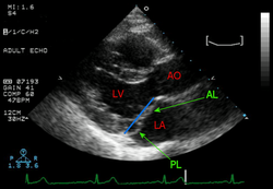
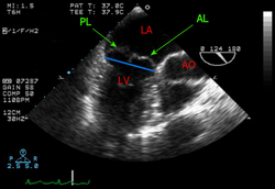
Transthoracic and transesophageal echocardiograms of mitral valve prolapse | |
Echocardiography, a noninvasive method of visualizing the heart, is the most useful method of diagnosing a prolapsed mitral valve. Two- and three-dimensional echocardiography are particularly valuable as they allow visualization of the mitral leaflets relative to the mitral annulus. This allows measurement of the leaflet thickness and their displacement relative to the annulus. Thickening of the mitral leaflets above 2 mm indicates mitral valve prolapse.
Treatment
Mitral valve prolapse can be treated with surgical replacement of the mitral valve. This may be necessary in as many as 11% of patients with classic MVP, and is indicated for patients with and ejection fraction below 60% and progressive left ventricular dysfunction.
Prevalence
Figures vary widely, but most recent studies of mitral valve prolapse indicate a prevalence of 1.3% for classic and 1.1% for nonclassic MVP. MVP occurs less frequently in children, and does not vary significantly with sex. Though the reasons are not understood, patients with mitral valve prolapse tend to be leaner with a relatively low body mass index.
References
- Mitral Valve Prolapse - Time For A Fresh Look (http://www.medreviews.com/pdfs/articles/RICM_22_73.pdf)
- Prevalence and Clinical Outcome of Mitral-Valve Prolapse (http://content.nejm.org/cgi/content/full/341/1/1)
- Mitral Valve Prolapse Prevalence and Complications (http://circ.ahajournals.org/cgi/content/full/106/11/1305)
- MedlinePlus Medical Encyclopedia: Mitral Valve Prolapse (http://www.nlm.nih.gov/medlineplus/ency/article/000180.htm)
External links
- Mitral Valve Prolapse - Texas Heart Institute Information Center (http://www.tmc.edu/thi/mvp.html)
- Mitral Valve Prolapse - Conscious Choice (http://www.consciouschoice.com/holisticmd/hmd093.html)
