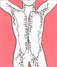
Lymphatic system
|
|
In mammals including humans, the lymphatic vessels (or lymphatics) are a network of thin tubes that branch, like blood vessels, into tissues throughout the body. Lymphatic vessels carry lymph, a colorless, watery fluid originating from interstitial fluid (fluid in the tissues). The lymphatic system transports infection-fighting cells called lymphocytes, is involved in the removal of foreign matter and cell debris by phagocytes and is part of the body's immune system. It also transports fats from the small intestine to the blood.
| Contents |
Composition of lymph
Lymph vessels are usually associated with the circulatory system vessels. Larger lymph vessels are alike veins. Lymph originates as blood plasma lost from the capillary beds of the circulatory system, which leaks out into the surrounding tissues. Although capillaries lose only about 1% of the volume of the fluid that passes through them, so much blood circulates that the cumulative fluid loss in the average human body is about 3L per day. The lymphatic system collects this fluid by diffusion into lymph capillaries, and returns it to the circulatory system. Once within the lymphatic system the fluid is called lymph, and has almost the same composition as the original interstitial fluid.
Lymphatic circulation
Unlike the circulatory system, the lymphatic system is not closed and has no central pump; the lymph moves slowly and under low pressure. Like veins, lymph vessels have one-way valves and depend mainly on the movement of skeletal muscles to squeeze fluid through them. Rhythmic contraction of the vessel walls may also help draw fluid into the lymphatic capillaries. This fluid is then transported to progressively larger lymphatic vessels culminating in the right lymphatic duct (for lymph from the right upper body) and the thoracic duct (for the rest of the body); these ducts drain into the circulatory system at the right and left subclavian veins (sub = underneath, ,clavian = collar bones i.e. they run along the underside of the collar bones).
Lymph vessels are present in the lining of the gastrointestinal tract. Whilst most other nutrients absorbed by the small intestine are passed on to the portal venous system to drain, via the portal vein, into the liver for processing, fats are passed on to the lymphatic system, to be transported to the blood circulation via the thoracic duct. The enriched lymph originating in the lymphatics of the small intestine is called chyle. The nutrients that are released to the circulatory system are processed by the liver, having passed through the systemic circulation.
Primary lymphoid organs
The thymus and bone marrow are the primary lymphatic organs. Lymphocytes are produced by stem cells in the bone marrow and then migrate to either the thymus or bone marrow where they mature. T-lymphocytes undergo maturation in the thymus (hence their name), and B-lymphocytes undergo maturation in the bone marrow. After maturation, both B- and T-lymphocytes circulate in the lymph and accumulate in secondary lymphoid organs, where they await recognition of antigens.
Secondary lymphoid organs
The spleen, lymph nodes, and accessory lymphoid tissue (including the tonsils and appendix) are the secondary lymphoid organs. These organs contain a scaffolding that support circulating B- and T-lymphocytes and other immune cells like macrophages and dendritic cells. When microorganisms invade the body or the body encounters other antigens (such as pollen), the antigens are transported from the tissue to the lymph. The lymph is carried in the lymph vessels to regional lymph nodes. In the lymph nodes, the macrophages and dendritic cells phagocytose the antigens, process them, and present the antigens to lymphocytes, which can then start producing antibodies or serve as memory cells to recognize the antigens again in the future.
The spleen contains lymphocytes that filter the blood stream rather than the lymphatics. Thus, the spleen has importance in fighting infections that have invaded the blood.
Accessory lymphoid tissues act as barriers along points of entry for infections, such as the lung, the reproductive system, and the gut. (See separate section below)
Lymph nodes
- See also: lymph node.
Along this network of vessels are small organs called lymph nodes. Clusters of lymph nodes are found in the underarms, groin, neck, chest, and abdomen. Lymph nodes act as filters, with an internal honeycomb of connective tissue filled with lymphocytes that collect and destroy bacteria and viruses. When the body is fighting an infection, these lymphocytes multiply rapidly and produce a characteristic swelling of the lymph nodes. Approximately 25 billion different lymphocytes migrate through each lymph node every day.
Accessory lymphoid tissue
Accessory lymphoid tissue consists of unorganized patches of lymphoid tissue found diffusely in various sites of the body. They are similar in function to the lymph nodes but are anatomically different and are sometimes termed extra-nodal lymphoid tissue. Tonsils and the vermiform appendix are examples of accessory lymphoid tissue.
Mucosa-associated lymphoid tissue (MALT) is the diffuse system of small concentrations of lymphoid tissue found in various sites of the body such as the gastrointestinal tract, thyroid, breast, lung, salivary glands, eye, and skin. The components of MALT are sometimes subdivided into GALT (gut or GI-associated lymphoid tissue), BALT (bronchial-associated lymphoid tissue), NALT (nose-associated lymphoid tissue), and SALT (skin-associated lymphoid tissue). A newly recognized entity is vascular-associated lymphoid tissue (VALT) that exists inside arteries; its role in the immune response is unknown.
Peyer's patches are a component of GALT found in the lining of the small intestines.

