Intermediate filament
|
|
Intermediate filaments are one component of the cytoskeleton - important structural components of living cells. Their size is intermediate between that of microfilaments and microtubules. They are assembled from several different proteins. IFs crisscross the cytosol from the nuclear envelope to the cell membrane.
| Contents |
Types
The different kinds of IFs share some basic characteristics: they are from 9 to 11 nm. in diameter and are very stable; their main function being a structural one. Different types of IFs are distinguished by the protein each is made of.
Lamin IFs
These form a network, the nuclear lamina, that supports the nuclear envelope. There are lamin A, B, and C filaments. Lamin A and C form the lamin network and are attached to the nuclear membrane by lamin B.
Keratin IFs
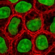
These proteins are the most diverse among IFs. The many isoforms are divided in two groups: "soft" keratins (cytokeratins) in epithelial cells (image to right), and "hard" keratins (hair keratins) which make up hair, nails, horns and reptilian scales. Regardless of the group, keratin can be acidic or basic. Acidic and basic keratins can bind each other to form acidic-basic heterodimers, these heterodimers can then associate to make a keratin filament.
Type III IFs
- Desmin IFs are structural components of the sarcomeres in muscle cells.
- Vimentin IFs can be found in fibroblasts and endothelial cells, they support the cell membrane and keep some organelles in a fixed place within the cytoplasm.
- Peripherin found in peripheral neurons.
- GFAP (glial fibrillary acidic protein) in glial cells.
Neurofilaments
These are found in nerve cells and are implicated in the radial growth of the axon.
- α-Internexin
- Neurofilament-L (NF-L)
- Neurofilament-M (NF-M)
- Neurofilament-H (NF-H)
Nestin
Intermediate filament type VI. It is found in neural stem cells.
Cell adhesion
At the plasma membrane, IFs are attached by adapter proteins forming desmosomes (cell-cell adhesion) and hemidesmosomes (cell-matrix adhesion).

