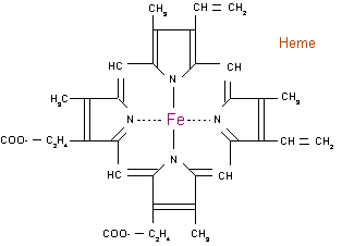
Heme
|
|
| Contents |
Types
There are three biologically important kinds of heme. The most common type of heme is called heme b; the others are heme a and heme c.
- Heme a differs from heme b in that a methyl side chain is oxidized into a formyl group, and one of the vinyl side chains has been replaced by an isoprenoid chain. Like heme b, heme a is not covalently bound to the apoprotein in which it is found. An example of a heme-containing protein that has heme a is cytochrome c oxidase.
- Heme b is, like heme a, not bound covalently to its apoprotein. It is the most abundant heme: both hemoglobin and myoglobin are examples of proteins that contain heme b.
- Heme c differs from heme b in that the two vinyl side chains are covalently bound to the protein itself. Examples of proteins that contain a c type heme are cytochrome c and the bc1 complex.
The names of cytochromes tend to (but not always) reflect the kinds of hemes they contain. Therefore, cytochrome a contains a a type heme, cytochrome c contains a c type heme, etc.
Function
The main function of heme is the retention of O2 and delivering it for enzymatic reactions. As the main site of oxidation in the cell is the mitochondrion, many heme-containing enzymes are located there.
Hemoglobin is not an enzyme, but a transporter. It binds oxygen in the pulmonary vasculature, where the pH is high and the pCO2, and releases it in the tissues, where the situations are reversed. The main mechanism behind this phenomenon is the steric organisation of the globin chain; a histidine residue, located adjacent to the heme group, becomes positively charged under acid circumstances, sterically releasing oxygen from the heme group.
Synthesis
Details of heme synthesis can be found in porphyrin.
Heme_synthesis.png
The pathway is initiated by the synthesis of D-Aminolevulinic acid (dALA or δALA) from the amino acid glycine and succinyl-CoA from the citric acid cycle (Krebs cycle). The rate-limiting enzyme responsible for this reaction, ALA synthase, is strictly regulated by intracellular iron levels and heme concentration. A low iron level, e.g. in iron deficiency, leads to decreased porphyrin synthesis, which prevents accumulation of the toxic intermediates. This mechanism is of therapeutic importance: infusion of heme arginate of hematin can abort attacks of porphyria in patients with an inborn error of metabolism of this process, by reducing transcription of ALA synthase.
The organs mainly involved in heme synthesis are the liver and the bone marrow, although every cell requires heme to function properly.

