Electrical conduction system of the heart
|
|
The normal electrical conduction in the heart allows the impulse that is generated by the sinoatrial node (SA node) of the heart to be propagated to (and stimulate) the myocardium (muscle of the heart). When the myocardium is stimulated, it contracts. It is the ordered stimulation of the myocardium that allows efficient contraction of the heart, therby allowing blood to be pumped to the body.
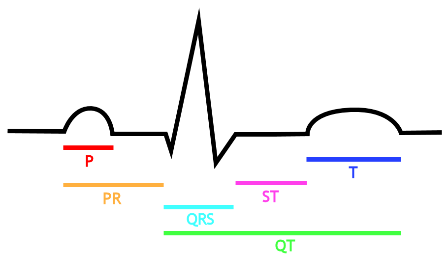
Under normal conditions, electrical activity is spontaneously generated by the SA node. This electrical impulse is propagated throughout the right and left atria, stimulating the myocardium of the atria to contract. The conduction of the electrical impulse throughout the atria is seen on the EKG as the P wave.
Electrical activity that originates from the SA node at a rate of between 50 beats/minute and 100 beats/minute is known as normal sinus rhythm. If a rhythm originating from the sinus node at a rate of less than 50 beats/minute, this is known as sinus bradycardia. On the other hand, if a rhythm originates from the sinus node at a rate of more than 100 beats/minute, this is known as sinus tachycardia.
As the electrical activity is spreading throughout the atria, it travels via specialized pathways from the SA node to the AV node, known as internodal tracts.
The AV node functions as a critical delay in the conduction system. Without this delay, the atria and ventricles will contract at the same time, and blood won't flow effectively from the atria to the ventricles. The delay in the AV node forms much of the PR interval on the EKG.
The distal portion of the AV node is known as the Bundle of His. The Bundle of His splits into two branches in the interventricular septum, the left bundle branch and the right bundle branch. The left bundle branch activates the left ventricle, while the right bundle branch activates the right ventricle. The left bundle branch is short, splitting into the left anterior fascicle and the left posterior fascicle. The left posterior fascicle is relative short and broad, with dual blood supply, making it particularly resistant to ischemic damage.
The two bundle branches taper out to produce numerous Purkinje fibers, which stimulate individual groups of myocardial cells to contract.
The spread of electrical activity through the ventricular myocardium produces the QRS complex on the EKG.

