
Image:FMRI.jpg
|
|
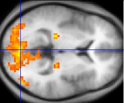
Sample fMRI data
This example of fMRI data shows regions of activation including primary visual cortex (V1, BA17), extrastriate visual cortex and lateral geniculate body in a comparison between a task involving a complex moving visual stimulus and rest condition (viewing a black screen). The activations (yellow-red) are shown (as is typical) against a background based on the average structural images from the subjects in the experiment.
Copyright info
Public-domain: copyright disclaimed Washington irving 07:49, 8 Mar 2004 (UTC)
| Missing image PD-icon.png Public domain | This image has been released into the public domain by the copyright holder, its copyright has expired, or it is ineligible for copyright. This applies worldwide.
|
File links
There are no pages that link to this file.